Another example of a visualization project done for Prof. Janos Vörös.
3-D graphics were done by Katja Messora. i was helping designing and planning the graphics and acted as a translater for the different languages of a scientist and a multimedia designer.
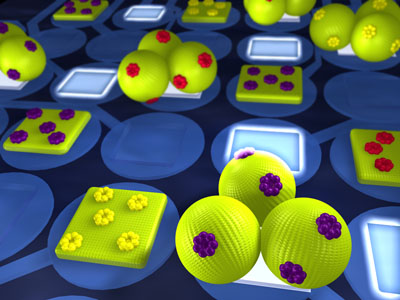
The image shows an array of electrodes coated with a functional polymer. It allows to address the single electrodes individually and in a second step to adsorb different biological units such as lipid vesicles on each electrode. This can be used in future technologies for the detection of diseases and testing of new drugs or to interface biology, such as nerve cells, with electronics.
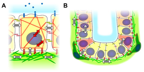
Here is an image which shows a simplified sketch of the different cues that influence cell behavior in a 3-dimensional context. I made this for a publication in Biointerphases published in 2006.
left: a single cell in contact with neighboring cells. An engineering approach has been used to simplify the image, simple symbols to show forces and mechanical interaction are emphasized.
right: this shows the same idea of dimensionality in a multicellular context. as an example a stem cell niche is shown. again the 3-D interaction of forces and architecture is highlighted.
Here is an animation of a single cell inside a square shaped microwell. This was part of my PhD work, for more infos about that check my CV on my site www.dusseiller.ch/cv
This was recorded using confocal laser scanning microscopy and the the 3D data was animated using bitplane Imaris.
The goal of the research project was to create 3-dimensional environments for single cells, and thus controlling the shape of the cells. The final goal was to demonstrate that the dimensionality is a crucial cue to influence cell behaviour. A fact that was highly neglected in standard cell culture. To visualize our point i spent a lot of time on new methods to analyze and show our findings using a variety of 3-d tools.
This example shows a epithelial cell grown inside a square microwell of approximately 20 microns width and 10 microns depth. The shape of the cell is highly influenced by the 3-D context and very different from spread cells found on hard and stiff petridishes. The cell membrane was fluorescently labeled and could thus be recorded in microscopy.