I am generally interested in a transdisciplinary approach to interface Science, Art and Education. I started my interdisciplinary work during my PhD at ETH Zürich bridging engineering concepts with biology and medicine.
My current projects are described on this site www.dusseiller.ch/labs
they include experimental electronics for musical instruments, communication in science, human machine interactions and other projects interfacing technology, science, arts and education.
Keywords
cell-based sensors, micro-3D cell culture, mechanotransduction, microfluidics, μTAS, tissue engineering, biointerfaces, hybrid devices, nanomedicine, ethics
Publications
My PhD thesis can be openly accessed on ETH's e-collection.
For a list of publications search for "dusseiller" on www.scholar.google.com, where you should find the up-to-date list of my patents and papers in journals such as Lab-on-a-Chip, Biomaterials and Biointerphases.
Research at ETH Zürich 2000 - 2007
The goal of the research project was to create 3-dimensional environments for single cells, and thus controlling the shape of the cells. The final goal was to demonstrate that the dimensionality is a crucial cue to influence cell behaviour. A fact that was highly neglected in standard cell culture. To visualize our point I spent a lot of time on new methods to analyze and show our findings using a variety of 3-d tools. For a more detailed overview of my former research @ ETH Zürich visit my former site on www.mat.ethz.ch.
Here is an image which shows a simplified sketch of the different cues that influence cell behavior in a 3-dimensional context. I made this for a publication in Biointerphases.
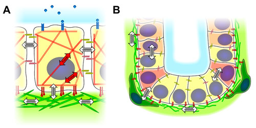
left: a single cell in contact with neighboring cells. An engineering approach has been used to simplify the image, simple symbols to show forces and mechanical interaction are emphasized.
right: this shows the same idea of dimensionality in a multicellular context. as an example a stem cell niche is shown. again the 3-D interaction of forces and architecture is highlighted.
Videos from my thesis "Micro- and Nanoengineering the 3-Dimensional Environment of Cells in Culture". Here is an animation of a single cell inside a square shaped microwell. This example shows a epithelial cell grown inside a square microwell of approximately 20 microns width and 10 microns depth. The shape of the cell is highly influenced by the 3-D context and very different from spread cells found on hard and stiff petridishes. The cell membrane was fluorescently labeled and could thus be recorded in microscopy.
This was recorded using confocal laser scanning microscopy and the the 3D data was animated using bitplane Imaris.
3-D reconstruction of CLSM stack of stably transfected (YFP-plasma membrane) MDCK cells cultured 4 h on a PS microwell array, circular microwells of 15 μm diameter. Most wells are filled by a single cell. No spreading of the cells on the plateau surface was observed. In the back corner a well seems to be occupied by two cells. Grid spacing 20 μm.